
Case Study
Automatic motion correction in life science

Darius Rückert
- last updated on Mar 10, 2025
Our study, conducted in collaboration with Kuwait University, focuses on reconstructing rabbit bone samples to investigate artificial bone growth and regeneration using different substrates. To assess bone formation, we scanned the samples using a CT scanner, measuring the amount of newly grown bone for each substrate.
Achieving high-quality images required long scan times, during which some water evaporated from the samples. This evaporation led to deformation, which standard CT reconstruction methods could not correct, resulting in significant artifacts, as seen in the images below.
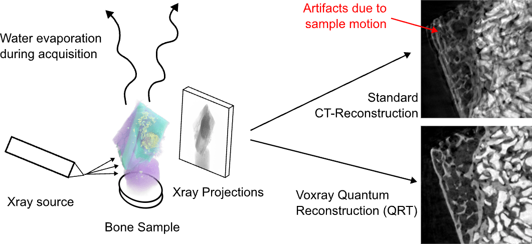
Water evaporation during the CT measurement caused deformation (motion) in the scanned sample. Using the Voxray QRT, the sample motion can be corrected resulting in a high-quality image.
To compensate for deformations caused by water evaporation during the scanning process, we apply our Quantum Reconstruction Technique (QRT). This method enables us to correct the geometry of each projection individually, effectively tracking and correcting the sample’s deformation.
Results
As a result, we achieve a highly accurate CT reconstruction, as if no deformation had occurred. This allowed the researcher to precisely evaluate bone growth without the need for rescanning, saving significant time and effort in the process.
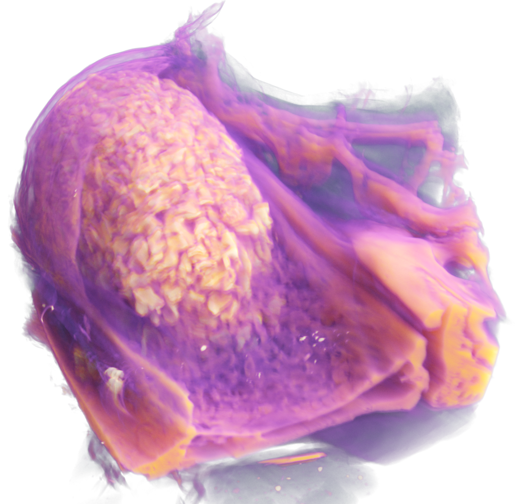

3D rendering of the bone sample. Standard CT reconstruction (left) and Voxray QRT (right)

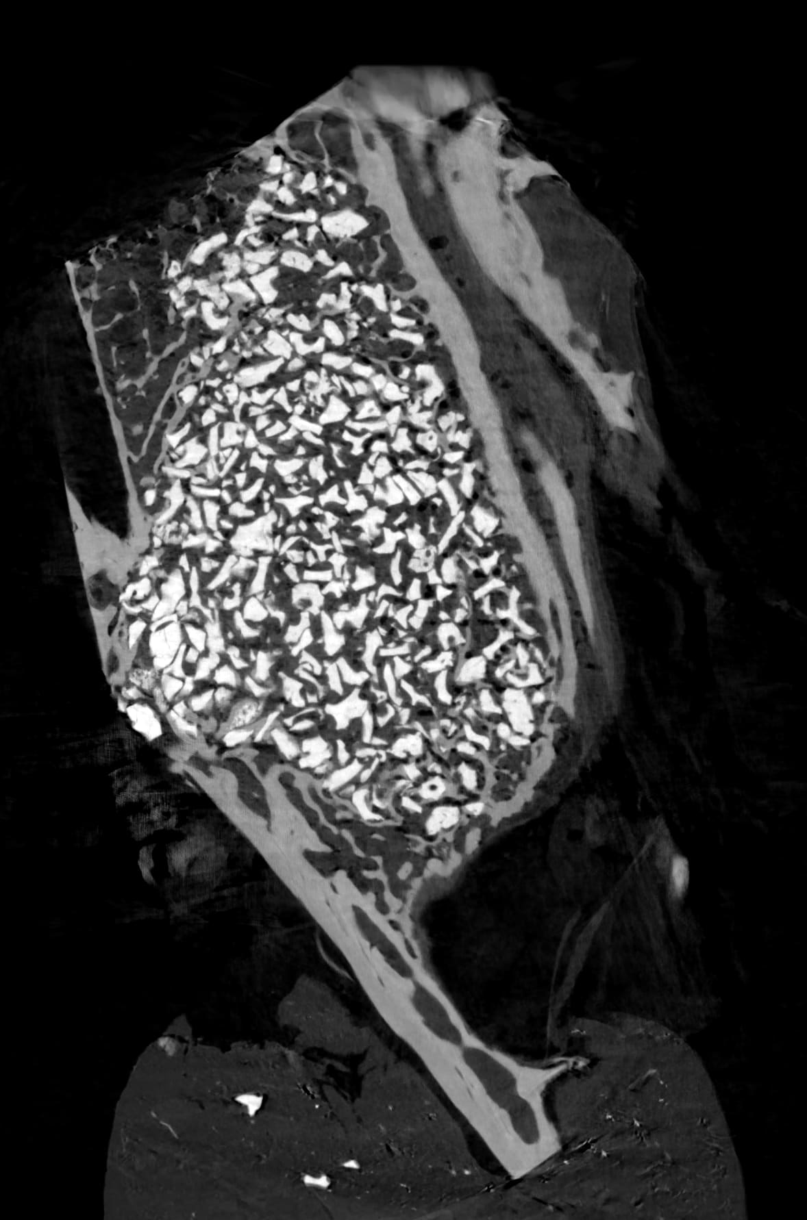
2D slice comparison of the standard CT reconstruction (left) and the Voxray QRT (right)

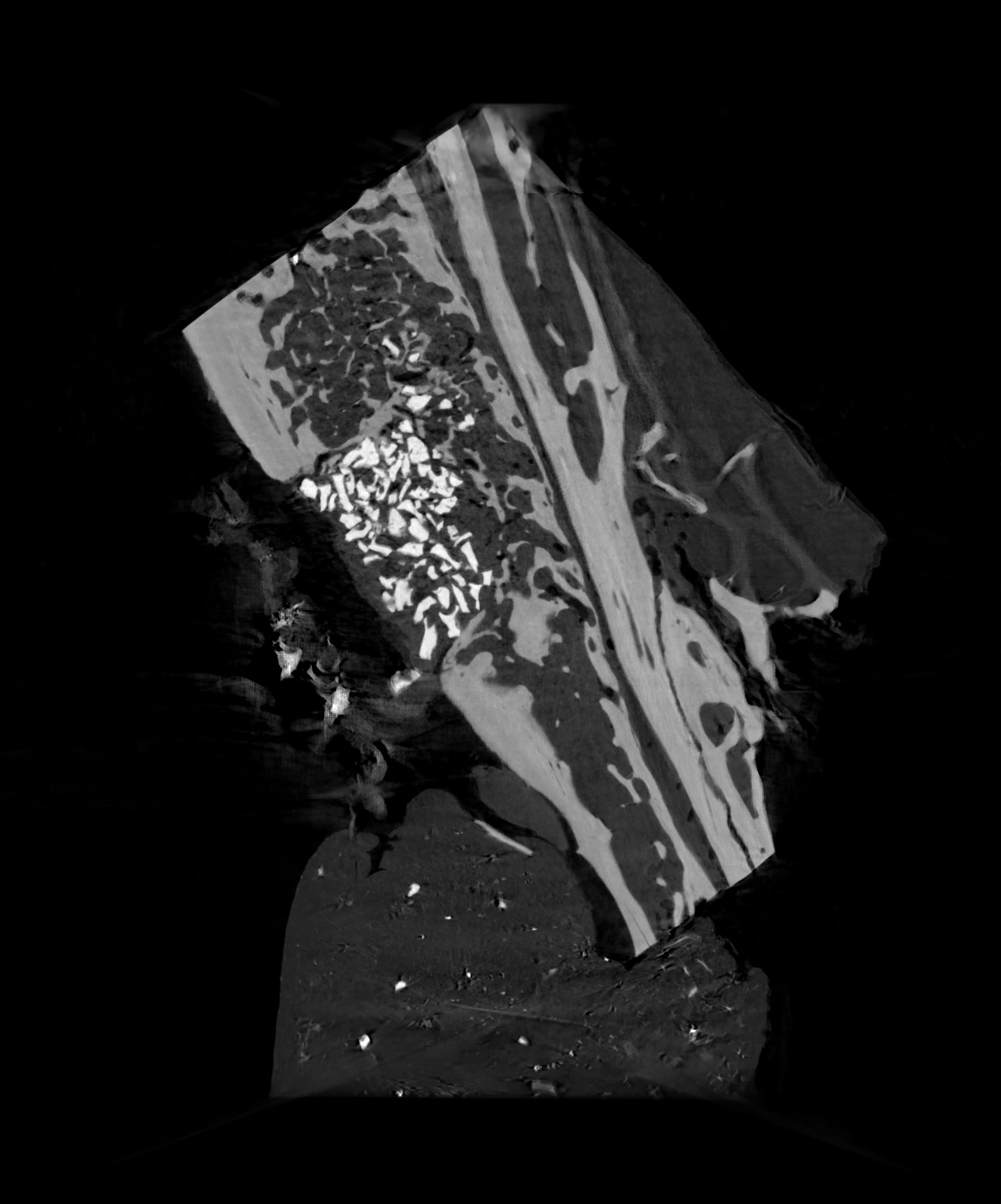
2D slice comparison of the standard CT reconstruction (left) and the Voxray QRT (right)
In this article